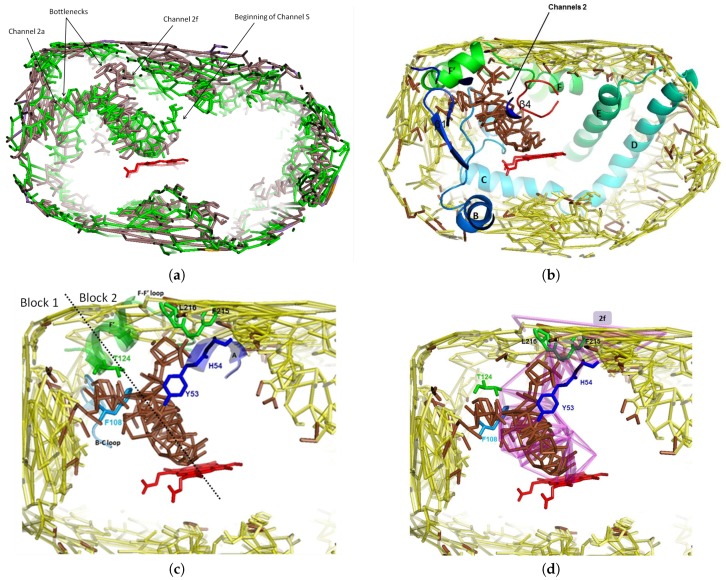
Figure 2.
Four representations of the main channel through its facial graph. The target atom is the iron of the heme group (in red). (a) Facial graph of the main channel to the active site of CYP3A4 computed from 2V0M (in brown, for CV = 7 Å), superposed to the facial graph of this channel computed from 1TQN (in green, for CV = 6 Å). The two facial graphs show the way in channel 2a: the one of 2V0M is the beginning of channel 2f with a bottleneck at its entry (not connected to the exterior); the two facial graphs show also the beginning of channels S. (b) The funnel shaped channel leading to the active site, computed from 2V0M, appearing at CV = 7 Å (in brown) superposed on the one computed at CV = 7.25 Å (in yellow); the part of the facial graph of channels 2 lies within sheet and C-terminal loop and B-C and F-G loops; only surface pockets were visible for CV > CV (in yellow), thus the evidence of a bottleneck at the entry of the two pathways. (c) Zoom of (b); bottleneck toward 2a: Phe108 (in B-C loop) and Thr224 (in helix F’); bottleneck toward 2f: Tyr53 and His54 (both in helix A), Phe215 and Leu216 (both in F-F’ loop); the motions of these residues induce the opening/closing of the bottlenecks; the residues constituting the bottleneck are in green and blue sticks; the dashed line separates blocks 1 and 2. (d) Zoom of (b), superposed on channel 2f computed at CV = 5.75 Å; six residues border the bottlenecks of channel 2f: four are located at the entry (in helix A and FF’ loop), Phe108 in the common part of 2a and 2f, and Thr224 where 2a and 2f separate.

