Figure 1.

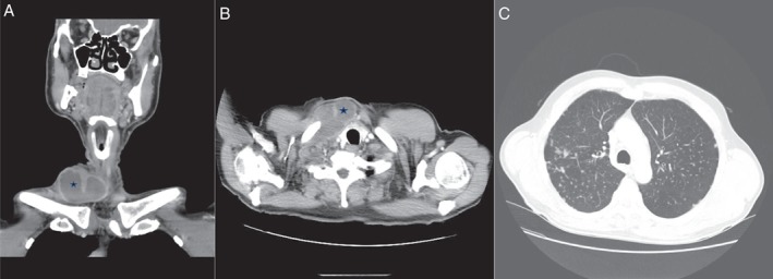
(A,B) Axial and coronal computed tomography (CT) scans of right lower neck cystic mass with peripheral enhancement. (C) CT thorax showing multiple well‐defined pulmonary nodules in bilateral upper lobe predominance.

(A,B) Axial and coronal computed tomography (CT) scans of right lower neck cystic mass with peripheral enhancement. (C) CT thorax showing multiple well‐defined pulmonary nodules in bilateral upper lobe predominance.