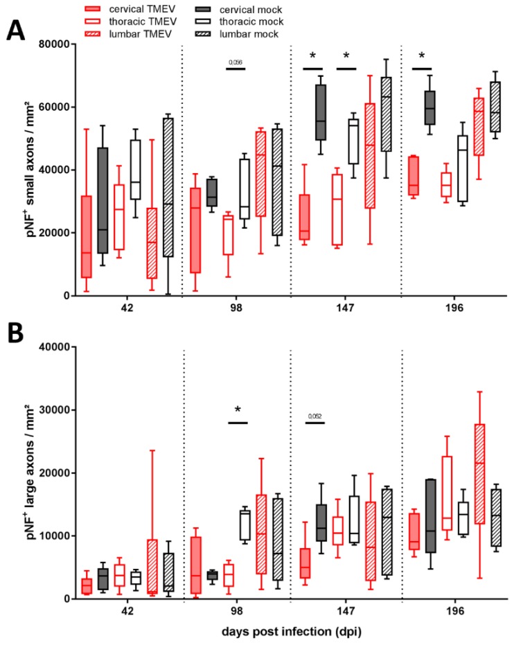
Figure 4.
Evaluation of axonal loss showed a decreased number of phosphorylated neurofilaments (pNF) positive small (1–4<μm) diameter axons within the thoracic and cervical spinal cord at 147 dpi and within the cervical segment at 196 dpi (A). Large (≥4 μm) axons were significantly decreased within the thoracic segment at 98 dpi (B). No significant differences between TMEV-infected groups comparing 147 and 196 dpi were detected. Graphs display box and whisker plots. Significant differences between groups detected by Mann–Whitney U-test were indicated by asterisks (* p ≤ 0.05).

