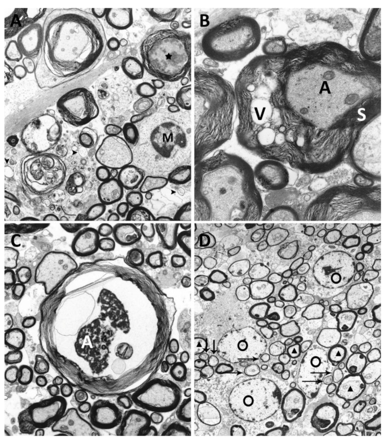
Figure 5.
Ultrastructural analysis of degenerative changes of axons and myelin sheaths of TMEV infected SJL-mice. (A) Demyelinated area with denuded axons [arrowheads] and infiltration of microglia/macrophages [M] and axonal degeneration [asterisk] within the ventrolateral areas of the spinal cord (TMEV infected animal, 42 dpi, magnification 5000×). (B) Vacuolation [V] of myelin sheath [S] surrounding an intact axon [A] (TMEV infected animal, 98 dpi, magnification 10,000×). (C) Axonal [A] degeneration characterized by shrinkage, an uneven axonal membrane and accumulation of electron dense material (TMEV infected animal, 98 dpi, magnification 5000×). (D) Multiple remyelinated axons [arrows], characterized by thinner myelin sheaths compared to normally myelinated fibers [triangles], characteristic for oligodendrocyte [O] mediated remyelination were detected (TMEV infected animal, 196 dpi, magnification 6300×).

