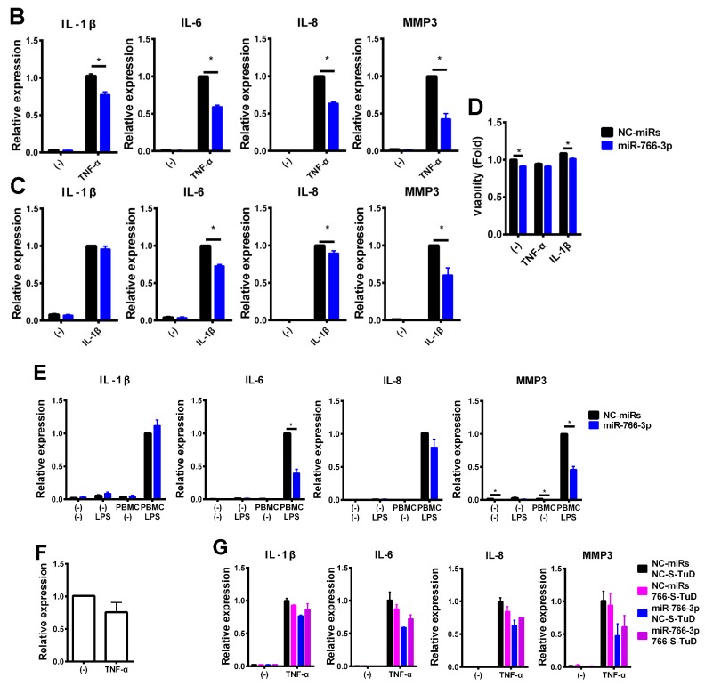
Figure 2.
Suppression of cytokine-induced inflammatory genes by miR-766-3p. (A) MH7A cells were transfected with the indicated concentrations of miR-766-3p mimic and exposed to TNF-α (10 ng/mL) for 24 h. The expression of IL-1β, IL-6, IL-8, and MMP3 was determined by a qPCR. (B–D) Cells were transfected with negative control miRs (NC-miRs) or miR-766-3p (5 nM). After incubation, cells were stimulated by (B,D) TNF-α or (C,D) IL-1β (10 ng/mL) for 24 h and subjected to (B,C) a qPCR or (D) a formazan assay. (E) miRNA mimic-transfected MH7A cells were co-cultured with peripheral blood mononuclear cells (PBMCs) using a Transwell system. Cells were treated with lipopolysaccharide (LPS; 1 μg/mL) for 24 h, and MH7A cells were subjected to a qPCR. The expression of the indicated genes was normalized to that of ACTB and then normalized to the respective values in TNF-α-, IL-1β- or PBMC + LPS-stimulated NC-miR-transfected cells. (F) MH7A cells were treated with TNF-α for 24 h. The expression of hsa-miR-766-3p was determined by a qPCR, and normalized to that of U6 small nuclear RNA. (G) MH7A cells were transfected with miRNA mimics. After incubation for 24 h, additional transfection with NC-S-TuD or 766-S-TuD (inhibitor of hsa-miR-766-3p), and after incubation for 24 h and treatment with TNF-α for 24 h. The cells were then subjected to a qPCR. Assays were performed in quadruplicate (A–E), or duplicate (F,G). Data are expressed as the mean ± SEM. Asterisks indicate statistically significant differences (p < 0.05).


