Figure 2.
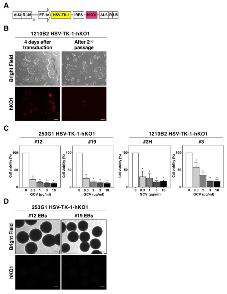
Silencing the transgene in human iPSCs. (A) Schematic representation of the integrated proviral form of the lentiviral vector expressing the HSV-TK-1 gene. HSV-TK-1, original HSV-TK gene; hKO1, humanized-codon Kusabira-Orange fluorescent protein gene. (B) Representative images of 1210B2 iPSCs 4 days after lentiviral transduction and after the second passage. Scale bar, 200 μm. (C) hKO1-positive iPSC clones, 253G1 HSV-TK-1-hKO1 (#12, #19) and 1210B2 HSV-TK-1-hKO1 (#2H, #3), were cultured in the presence of various concentrations of GCV for 3 days. Cell viability was assessed by the CCK-8 assay. The percent cell viability was calculated relative to cells in the absence of GCV. Data represent the mean ± SEM (n = 4). *, p < 0.05. (D) Representative images of EB formation of 253G1 HSV-TK-1-hKO1 iPSCs (#12, #19) on day 14. hKO1 fluorescence signal was not detected. Scale bar, 200 μm.

