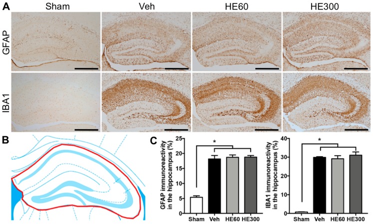
Figure 3.
Reactive gliosis after pilocarpine-induced SE was not altered by either low- or high-dose HE administration. (A) Representative photomicrographs showing immunohistochemistry to glial fibrillary acidic protein (GFAP) and ionized calcium-binding adapter molecule 1 (IBA1) in sham, vehicle (Veh)-, 60 mg/kg HE- (HE60), and 300 mg/kg HE-treated groups (HE300). Scale bar: 500 μm. (B) A drawing of the hippocampus indicating the region for the quantitative analysis of reactive gliosis (shown in red). (C) Graphs showing the immunoreactivity to GFAP and IBA1 in the hippocampus. Note that both GFAP and IBA1 immunoreactivity were significantly increased by SE, but there were no differences in the glial activation among Veh-, HE60-, and HE300-treated groups. Data are shown as mean ± SEM. n = 6 (sham), n = 10 (Veh), n = 8 (HE60), n = 6 (HE300). * p < 0.05; one-way ANOVA with Tukey’s post-hoc test.

