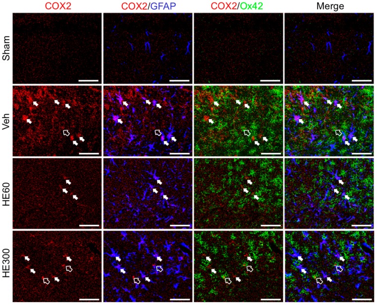
Figure 5.
HE treatment at 60 mg/kg suppressed hippocampal COX2-expressing glial cells after pilocarpine-induced SE. Triple immunohistochemistry for COX2 (red), GFAP (blue), and Ox42 (green) demonstrated that COX2-expressing cells in the Veh-treated group turned out to be GFAP-positive astrocytes (filled arrows) and Ox42-positive microglia (hollow arrow). In the group treated with 60 mg/kg HE, COX2/Ox42-coexpressing cells were not observed, and COX2/GFAP immunoreactivity was reduced. However, in the group treated with 300 mg/kg HE, COX2-expressing astrocytes and microglia were comparable with those in the Veh-treated group. Scale bar: 50 μm.

