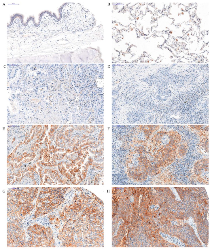
Figure 1.
Positive membranous immunohistochemical reaction (brown) indicating PD-L1 expression performed on healthy lung tissue (A,B) and in different grades of malignancy in adenocarcinoma (AC) (C,E,G) and squamous cell cancer (SCC) (D,F,H). Lack of PD-L1 expression—healthy lung tissue (A) and PD-L1 expression in macrophages—positive control (B). Expression of PD-L1 increased in higher malignancy grade in AC—G1 (C), G2 (E), and G3 (G), and in SCC—G1 (D), G2 (F) and G3 (H), magnification, ×200.

