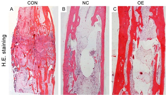
Figure 3.

(A-C) All samples from Group CON (A), Group NC (B) and Group OE (C) after 8-week consolidation were observed under a light microscope after H&E staining. The newly formed cortex in Group NC and Group OE was more continuously than in Group CON. In Group NC, the newly formed trabeculae in the distraction gap were thin, and partial trabeculae bridged discontinuously. More mature and regular trabecular bone were seen in Group OE.
