Figure 4.

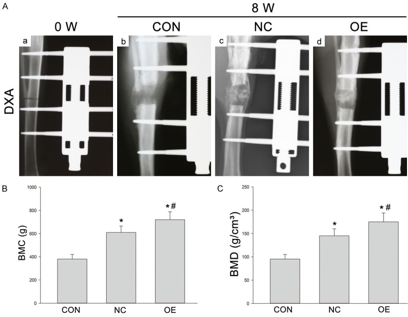
(A) X-ray examination of new bone formation in three groups after 8-week consolidation (A: b-d). Lateral radiographs of the distracted tibia before the distraction (0 week) (a). The newly regenerated bone in Group OE showed more mature and regular trabeculae as compared to other two groups. (B, C) BMC (g) and BMD (g/cm2) of the regenerated bone in Group CON, Group NC and Group OE after 8-week consolidation as measured by DXA. *P < 0.05 vs Group CON; #P < 0.05 vs Group NC.
