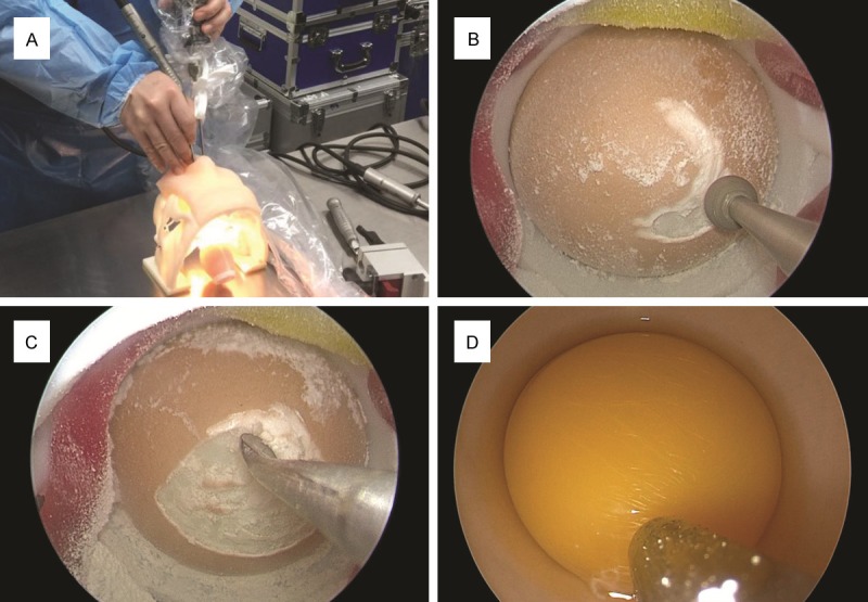
Figure 2.

Endoscopic endonasal surgical training with 3D printed model. (A) An egg was secured at the special sellar base. The endoscope and endoscopic tools were passed through the nostrils, respectively. (B-D) The tools were used to finish drilling of the shell (B), curetting and biting of the shell to further enlarge area of shell removal (C), and aspirating off egg white (D).
