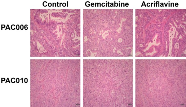
Figure 4.

Representative H&E stain of tumor tissues at the end of the treatment. Tumor tissue was obtained from the mice at the end of the treatment period or their respective controls. Routine H&E staining was performed and histology was visualized by microscope. Original magnification of histological images is × 20; scale bar 50 µm.
