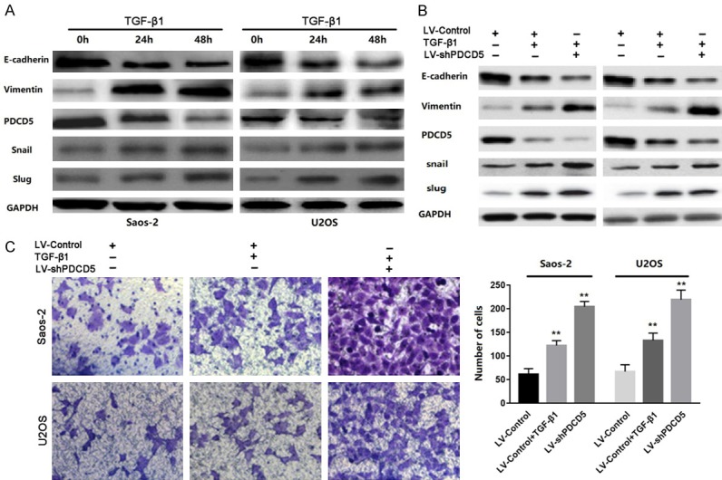
Figure 7.

PDCD5 participates in TGF-β1-induced EMT. A. Western blot of PDCD5, vimentin, E-cadherin, Snail and slug in the indicated cells in response to treatment with 10 ng/mL TGF-β1 for 0, 24, and 48 h. B. Twenty-four hours post-transfection of LV-Control or LV-shPDCD5 lentivirus, the cells were treated with TGF-β1 (2 ng/ml) for an additional 48 h. The expressions of PDCD5, E-cadherin, vimentin, Snail and slug were detected by Western blot. C. Representative images and data of a transwell invasion assay for Saos-2 and U2OS cells. TGF-β1 stimulation significantly increased the invasiveness of both tumor cell lines compared with that in the absence of TGF-β1 (LV-Control), and LV-shPDCD5 lentivirus cells enhanced the effect of TGF-β1. Each bar represents the mean ± SD, **P<0.01. All images are representative of three independent experiments with similar findings.
