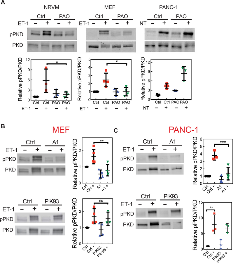
Fig. 7. PI4P depletion at the PM inhibits PKD activation.
(A) The indicated cells were treated with 10 μM PAO for 15 min to deplete global PI4P, which was followed by the addition of agonist for 30 min (100 nM ET-1 or 100 nM NT). The cells were then analyzed by Western blotting with antibodies against total PKD and pPKD (Ser916). (B) MEFs were treated with 100 nM A1 or 300 nM PIK93 for 15 min, which was followed by the addition of 100 nM ET-1 for 30 min. The cells were then analyzed by Western blotting with antibodies against total PKD and pPKD (Ser916). (C) PANC-1 cells were treated with A1 or PIK93 for 15 min, which was followed by the addition of 100 nM NT for 30 min. The cells were then analyzed by Western blotting with antibodies against total PKD and pPKD (Ser916). Western blots in all panels are representative of three independent experiments. Graphs in each panel show the abundance of pPKD relative to that of total PKD from three independent experiments. Data are means ± SEM and were analyzed by one-way ANOVA with Tukey’s post-test. *P < 0.05, **P < 0.01, and ***P < 0.001.

