
Figure 4.
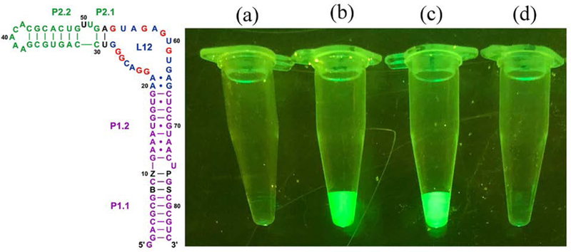
(Left) Schematic showing the full hachimoji spinach variant aptamer; additional nucleotide components of the hachimoji system are shown as black letters at positions 8, 10,76, and 78 (B, Z, P, and S respectively). The fluor binds in loop L12 (25). (Right) Fluorescence of various species in equal amounts as determined by UV. Fluorescence was visualized under a blue light (470 nm) with an amber (580 nm) filter. From left to right: (a) Control with fluor only, lacking RNA, (b) hachimoji spinach with the sequence shown in the left panel (c) native spinach aptamer with fluor, and (d) fluor and spinach aptamer containing Z at position 50, replacing A:U pair at positions 53:29 with G:C to restore the triple observed in the crystal structure. This places the quenching Z chromophore near the fluor; CD spectra suggest that this variant had the same fold as native spinach (Fig. S8).