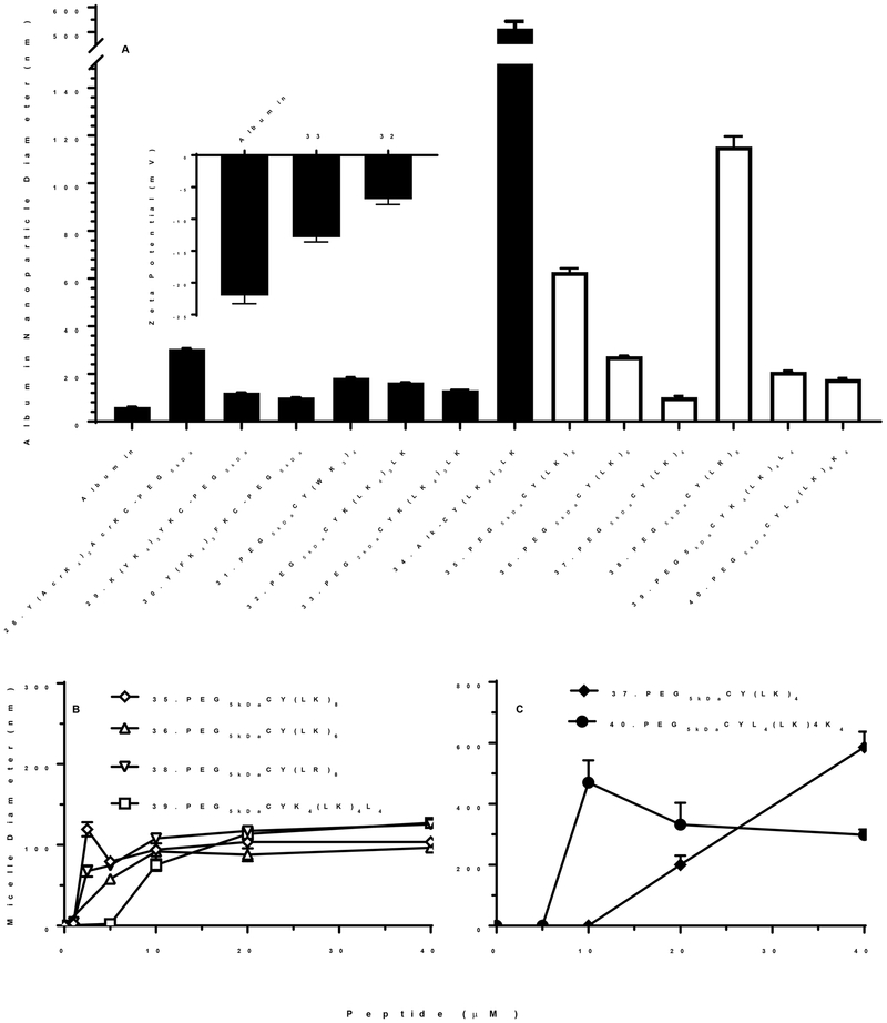
Figure 6. Particle Size Analysis of PEG-Peptide Albumin Nanoparticles and Micelles.
The particles size of albumin nanoparticles was determined by dynamic light scattering following the addition of 40 nmols of PEG-peptide with 5 mg of BSA in a total volume of 1 ml of 5 mM Hepes pH 7.4 (Panel A). The mean and standard error was determined from ten analysis. The albumin nanoparticle size ranged from 10–120 nm in diameter depending of PEG-peptide structure. Zeta potential of albumin nanoparticles prepared with 32 (PEG5kDa-Cys-Tyr-(Leu-Lys4)3-Leu-Lys) and 33 (PEG2kDa-Cys-Tyr-(Leu-Lys4)3-Leu-Lys) are compared to albumin (Panel A inset). PEG-peptides were independently analyzed for micelle formation by dynamic light scattering at increasing concentrations of 1–40 nmol in 1 ml of 150 mM sodium chloride, 50 mM sodium phosphate pH 7.4 (Panel B and C). PEG-peptide with a closed bar in panel A failed to form micelles whereas PEG-peptides with open bar are represented in panel B and C.

