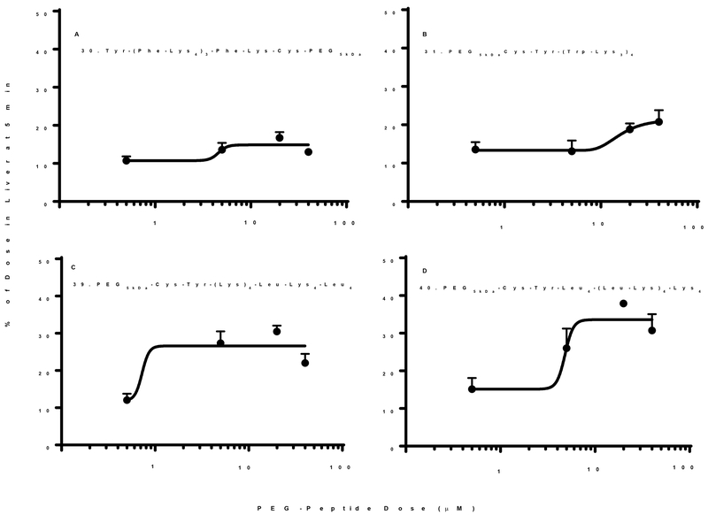
Figure 8. Biodistribution of Micelle Forming Low Molecular Weight PEG-Peptides.
The percent of 125I-PEG-peptide recovered from the liver at a biodistribution time of 5 min following i.v. administration of triplicate mice under dose escalation of 1–80 nmol is illustrated. Each PEG-peptide showed an increase in the percent of dose in liver upon dose escalation suggesting the formation of PEG-peptide micelles that failed to saturate liver uptake. However, only 39 and 40 produced measurable micelles (Fig. 6B,C). The curve fitting resulted in an R2 =0.5, 0.74, 0.76 and 0.78 for panels A-D. A value of p < 0.05 was determined for PEG-peptides 30, 31, 39, and 40 when comparing 1 and 80 nmol dose.

