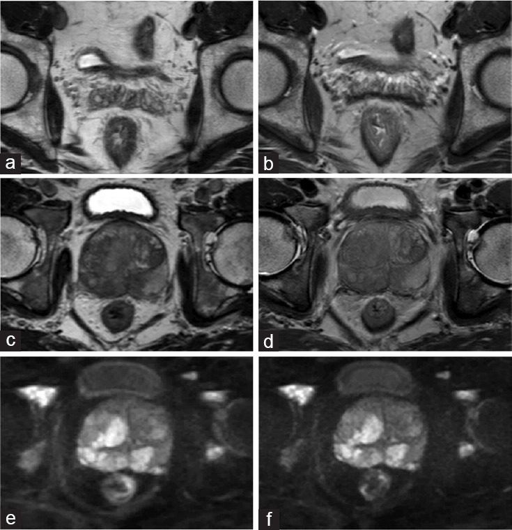
Figure 1.

(a) The image of MISS-T2WI of a 52-year-old man with prostate cancer. (b) The image of conventional T2WI of the same patient. The two figures showed the seminal vesicles delineation was better in MISS-T2WI than the conventional sequence. (c) 3D-T2WI in MISS of a 66-year-old man with prostate cancer. (d) 2D-T2WI in conventional T2WI of the same patient. The lesion showed slightly better lesion contrast on MISS-T2WI than the conventional T2WI. (e) DWI in MISS. (f) Conventional DWI. There was no obvious difference of image quality between MISS-DWI and conventional DWI. MISS: multiple instantaneous switchable scan; T2WI: T2-weighted imaging; 3D: three-dimensional; DWI: diffusion-weighted imaging.
