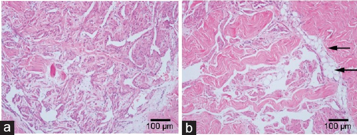
Figure 3.

The H and E staining of penile corpus cavernousum. Transverse sections of penile tissues at the borderline between skin and hair was fixed in 10% formalin buffer solution, embedded in paraffin, sectioned (6 μm) and stained with H and E. Compared with (a) control rabbits, the sinusoid lacunae of (b) hyperlipidemic rabbits were larger, and the arrangements of smooth muscle cells and fibrocytes were looser. Numerous empty cellular structures, probably adipocytes (arrow) were seen under the tunica albuginea in hyperlipidemia samples. Magnification ×100; scale bars = 100 μm.
