Figure 3.

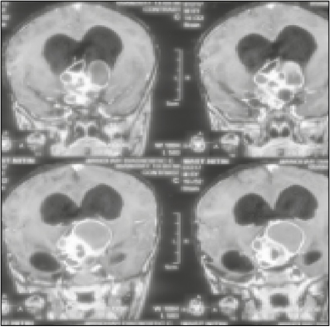
MRI of brain, contrast-enhanced, coronal section image showing giant mass lesion with epicenter in sellar with extension into parasellar and posterior fossa region

MRI of brain, contrast-enhanced, coronal section image showing giant mass lesion with epicenter in sellar with extension into parasellar and posterior fossa region