
Figure 1.
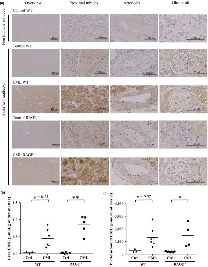
CML accumulation in the kidneys of WT and RAGE−/−mice was diet‐dependant. (a) Representative localization of protein‐bound CML studied by IHC on kidney sections showed that mice fed a CML‐enriched diet exhibited predominantly tubular staining. From left to right: low magnification, high magnification on proximal tubules, arterioles and glomeruli. (b‐c) Quantification by HPLC‐MS/MS of (b) free and (c) protein‐bound CML in kidneys. *p < 0.05, **p < 0.01, Kruskal–Wallis test