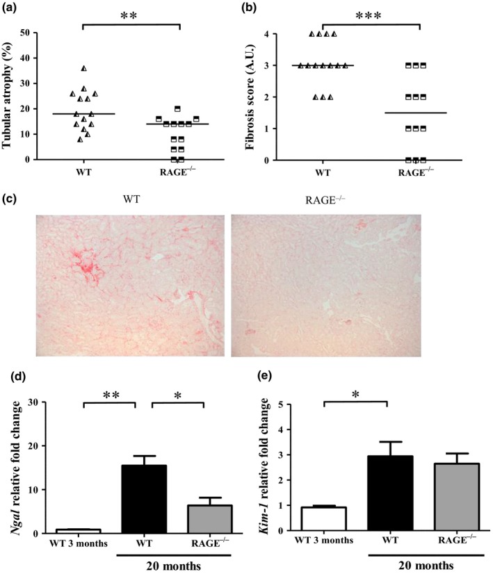
Figure 3.

RAGE−/− mice were protected against tubulo‐interstitial aging. Quantification of (a) tubular atrophy was determined by PAS staining and (b) interstitial fibrosis by Sirius red staining. (c) Representative Sirius red staining of control and CML WT and RAGE−/− mice at low magnification (x 100). (d‐e) Expression of kidney injury markers Ngal (d) and Kim‐1 (e) in renal tissue from 3‐ or 20‐month‐old WT and RAGE−/− mice (mean ±SEM, n = 3 for 3‐month‐old mice and n = 10 for 20‐month‐old mice). *p < 0.05, **p < 0.01, ***p < 0.001, unpaired t test or Kruskal–Wallis test for multiple comparisons
