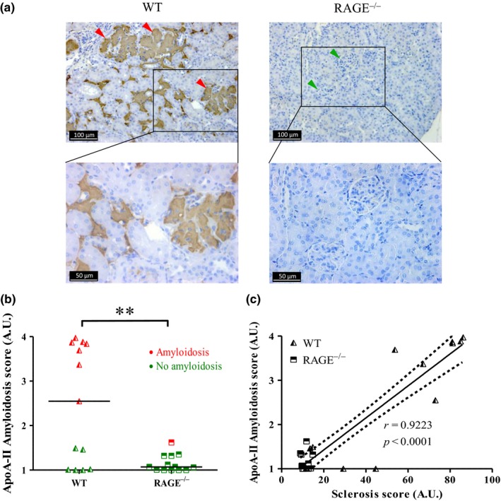
Figure 5.

AApoA‐II was largely absent in RAGE−/− mice. AApoA‐II in kidney tissue sections from 20‐month‐old WT mice (left panel) and RAGE−/− mice (right panel) were immunostained using an anti‐ApoA‐II antibody (a) green arrowheads, no deposits; red arrowheads, extensive deposits (representative image). (b) Scoring of ApoA‐II amyloid deposits obtained from IHC and calculated from the grading of 100 glomeruli per mouse, where 1 = no deposit, 2 = small, 3 = moderate and 4 = extensive deposits. Red dots, amyloidosis‐positive mice; green dots, amyloidosis‐negative mice. (c) Linear regression analysis between AApoA‐II and sclerosis scores obtained for all four conditions, y = 0.036 x + 0.70, r = 0.9223, dotted line: 95% confidence intervals (C). **p < 0.01, unpaired t test
