Figure 3.

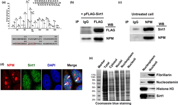
Physical interaction between Sirt1 with nucleophosmin (NPM). (a) Representative mass spectrogram of a peptide segment of NPM (sequence indicated by the red box below) co‐immunoprecipitated with Sirt1. (b) Immunoprecipitation (IP) of FLAG‐Sirt1 fusion protein in transfected cells and Western blot (WB) detection of bound NPM. Normal IgG was used in IP as a negative control. (c) Immunoprecipitation of endogenous NPM in untreated cells and WB detection of bound Sirt1. (d) Fluorescent confocal microscopic images showing partial co‐localization of NPM with Sirt1 (yellow color as indicated by the arrows) in the nucleolar compartment. DAPI was used to stain the nucleus. (e) Western blot detection of Sirt1 protein in purified nucleoli (right panel). Fibrillarin and nucleostemin were nucleolar markers. Histone H3 was a nuclear marker (which was also present in nucleoli to a lesser extent). The left panel showed a Coomassie blue‐stained PAGE gel showing the electrophoresis patterns of various subcellular fractions as indicated. All experiments were performed using HeLa cells
