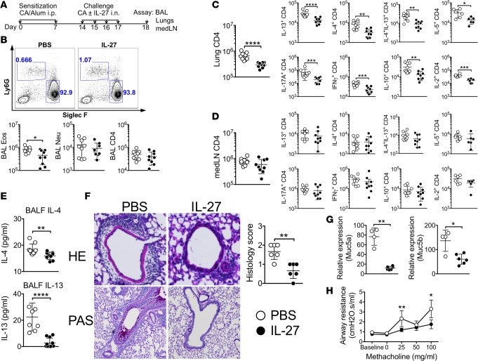
Figure 1. Intranasal IL-27 administration attenuates the development of allergic airway inflammation.
(A) Experimental protocol. B6 mice were injected i.p. on days 0 and 7 with CA in alum adjuvant. Starting at day 14, mice were intranasally challenged for 4 consecutive days with CA alone or together with IL-27. Mice were sacrificed 24 hours after the last challenge. (B) BAL cells were examined for Ly6G and Siglec F expression. Differential cell count was performed by FACS analysis. (C and D) Lung (C) and draining medLN (D) cells were harvested and stimulated ex vivo to assess intracellular cytokine expression. (E) IL-4 and IL-13 secretion in the BALF was determined using a CBA assay. (F) H&E and PAS staining of the lung tissues (original magnification ×20 and ×10, respectively) was used to evaluate inflammation. Histology score was determined as described in Methods. (G) Muc5a and Muc5b mRNA expression in the lung was determined by qPCR analysis. (H) Airway resistance was measured by flexivent experiments. Each symbol represents individually tested animal. The data shown are the mean ± SD of 3 independent experiments (n = 9). *P < 0.05, **P < 0.01, ***P < 0.001, ****P < 0.0001, as determined by Mann-Whitney nonparametric test. CA, cockroach antigen; BALF, bronchoalveolar lavage fluid; Eos, eosinophils; Neu, neutrophils; medLN, mediastinal lymph node.

