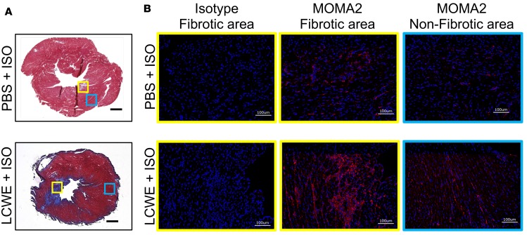
Figure 4. Increased myocardial macrophages infiltration in LCWE-induced KD vasculitis following ISO treatment.
WT mice were injected with either PBS or LCWE and allowed to recover for 5 weeks before receiving ISO for 10 consecutive days. Twenty-four hours after the last ISO injection, hearts were harvested and used for histological analysis. (A) Representative images of Masson’s trichrome–stained heart sections from PBS- or LCWE-injected KD mice treated with ISO. Scale bars: 500 μm. (B) MOMA2 immunofluorescent staining (red) in heart tissue fibrotic area (yellow boxes) and nonfibrotic area (blue boxes) from the mouse groups in A. DAPI (blue) was used to visualize nuclei. Scale bars: 100 μm. LCWE, Lactobacillus casei cell wall extract; ISO, isoproterenol.

