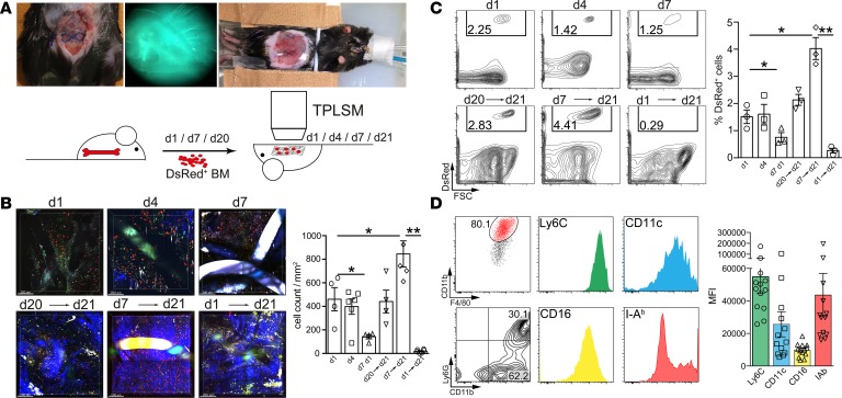
Figure 4. Intravital 2-photon laser-scanning microscopy imaging of abdominal meshes following adoptive Actin-DsRed+ BM cell transfer.
2 × 10⁷ BM cells from Actin-DsRed donor mice were transferred i.v. into mesh-implanted mice and followed by intravital 2-photon laser-scanning microscopy (TPLSM) imaging (imaging for up to 3 hours). (A) Representative images of the experimental setting and schematic overview of the experimental design. (B) TPLSM images of cellular infiltrates at early stages of foreign body reaction (top row). Cells were injected 12 hours following surgery and analyzed at day 1, day 4, and day 7 (acute FBR). Imaging at day 21 (bottom row) was commenced following cell injection 1, 14, and 20 days before imaging (chronic FBR). Images were composed of 4 adjacent view fields. Mesh fibers are visible as either black or highly fluorescent coherent structures. Blue fibers show collagen fibers visualized by second harmonic generation. Scale bar: 200 μm. Statistical analysis was performed by cell counting in at least 4 view fields of surrounding mesh fibers with a penetration depth of 200 μm. (C) Flow cytometric assessment of cellular infiltration visible in B. Cells were stained for CD45, Ly6G, CD11b, F4/80, CD11c, I-Ab, Ly6C, and CD16. Cells were gated as CD45+Ly6G–DsRed+ cells and for quantification of relative monocyte recruitment efficacy. (D) Flow cytometric subtyping of cellular infiltrates from DsRed+ cells after 14 days (day 7→day 21). Cells were pregated as CD45+Ly6G–DsRed+ and analyzed for their expression of the indicated surface markers. Expression strength was quantified by calculating the MFI of the indicated markers. Exemplary results are shown in the dot plots; statistical analysis was performed from ≥3 animals per group. Student’s t test: *P < 0.05; **P < 0.01. Error bars represent mean ± SD.

