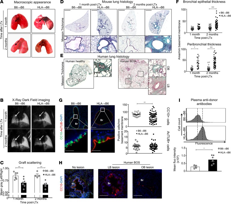
Figure 1. HLA-A2–knockin lung allografts are chronically rejected in a mouse model of orthotopic lung transplantation and present human-like signs of lymphocytic bronchiolitis.
Left lungs from C57BL/6J (B6) and HLA-A2–knockin (HLA) mice on a B6 background (HLA) were orthotopically transplanted into B6 recipients and analyzed 1 month (B6→B6, n = 4, HLA→B6, n = 4) and 2 months (B6→B6, n = 4, HLA→B6, n = 5) later. (A) Heart-lung blocks from the indicated mice. The arrows show the grafts. (B) Lungs acquired with the x-ray dark-field imaging technique. The arrows show the grafts. (C) Quantification of the left lung graft scattering. Data are expressed as mean ± SEM and were analyzed with a 2-way ANOVA with a Bonferroni post-test; **P < 0.01. (D) Scans (original magnification, ×2; scale bars: 1000 μm) and zoomed bronchi (original magnification, ×20; scale bars: 100 μm) from indicated transplanted mice stained with Masson’s trichrome. (E) Scans of Masson’s trichrome–stained explants from healthy and transplanted human lungs with bronchiolitis obliterans syndrome (BOS), with magnifications of bronchioles (LB, lymphocytic bronchiolitis; OB, obliterative bronchiolitis). (F) Quantification of the epithelial and peribronchial areas of the indicated mice. Data are expressed as mean ± SEM of all the quantified bronchi and analyzed with a 2-way ANOVA with a Bonferroni post-test; ***P < 0.001. (G) Double immunofluorescence and quantification of the CC10+ club cells and AcTUB+ ciliated cells. Scale bars: 100 μm (top); 200 μm (bottom). Data are expressed as mean ± SEM of all the quantified bronchi and were analyzed with a Mann-Whitney test. (H) Immunofluorescence from bronchioles of human explants stained with anti-CC10 (83) and counterstained with DAPI (blue). Scale bars: 100 μm. BOS, Bronchiolitis obliterans syndrome; LB, lymphocytic bronchiolitis; OB, obliterative bronchiolitis. (I) Flow cytometry of anti–HLA-A2 anti-donor antibody titers in the transplanted mice plasma, 2 months after LTx, and semiquantitative assessment of the anti-HLA Ab levels expressed as mean fluorescence intensity. Data are expressed as mean ± SEM and were analyzed with a Mann-Whitney test; *P < 0.05.

