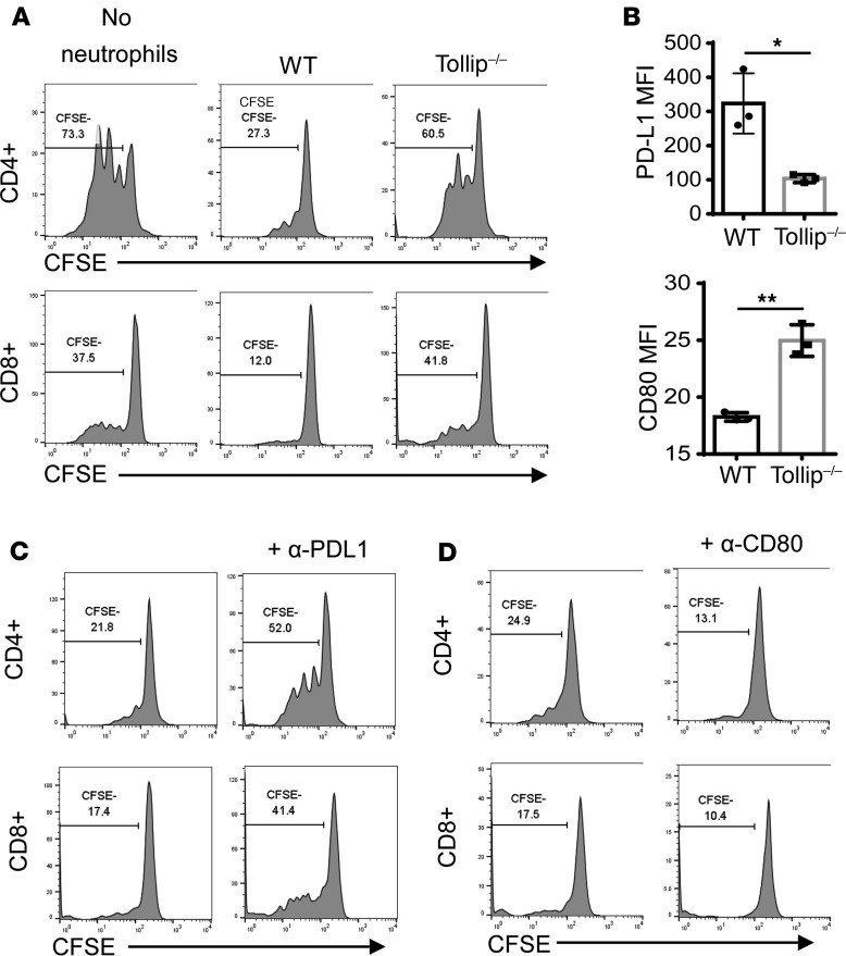
Figure 3. Tollip deficiency released the neutrophil suppression on T cell proliferation via PD-L1/CD80.
(A) CFSE-labeled splenocytes were cocultured with GM-CSF–primed neutrophils in anti-CD3 antibody–coated plates for 72 hours. Representative results are shown. (B) PD-L1 and CD80 expression on GM-CSF–primed neutrophils. Statistical significance compared with WT in the same treatment conditions was determined by Welch’s test. *P < 0.05, **P < 0.01. (C) In the presence of anti–PD-L1 antibody, CFSE-labeled splenocytes were cocultured with GM-CSF–primed WT neutrophils in anti-CD3 antibody–coated plates for 72 hours. (D) In the presence of anti-CD80 antibody, CFSE-labeled splenocytes were cocultured with GM-CSF–primed WT neutrophils in anti-CD3 antibody–coated plates for 72 hours.

