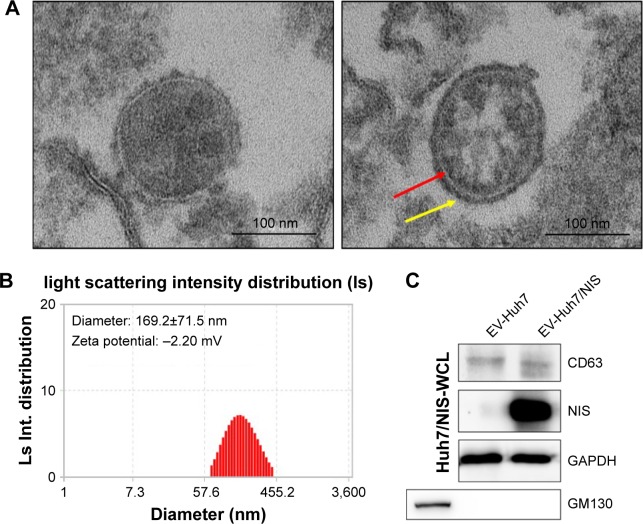
Figure 2.
Characterization of EVs isolated from Huh7/NIS.
Notes: (A) Morphology of EV-Huh7/NIS confirmed by transmission electron microscopy, arrow indicates the lipid bilayer (scale bar: 100 nm). (B) Size and Zeta potential of EV-Huh7/NIS determined by ELS (n=3; average diameter: 169.2±71.5 nm). (C) Western blot analysis of EV-Huh7/NIS and Huh7/NIS.
Abbreviations: EVs, extracellular vesicles; ELS, electrophoretic light scattering.

