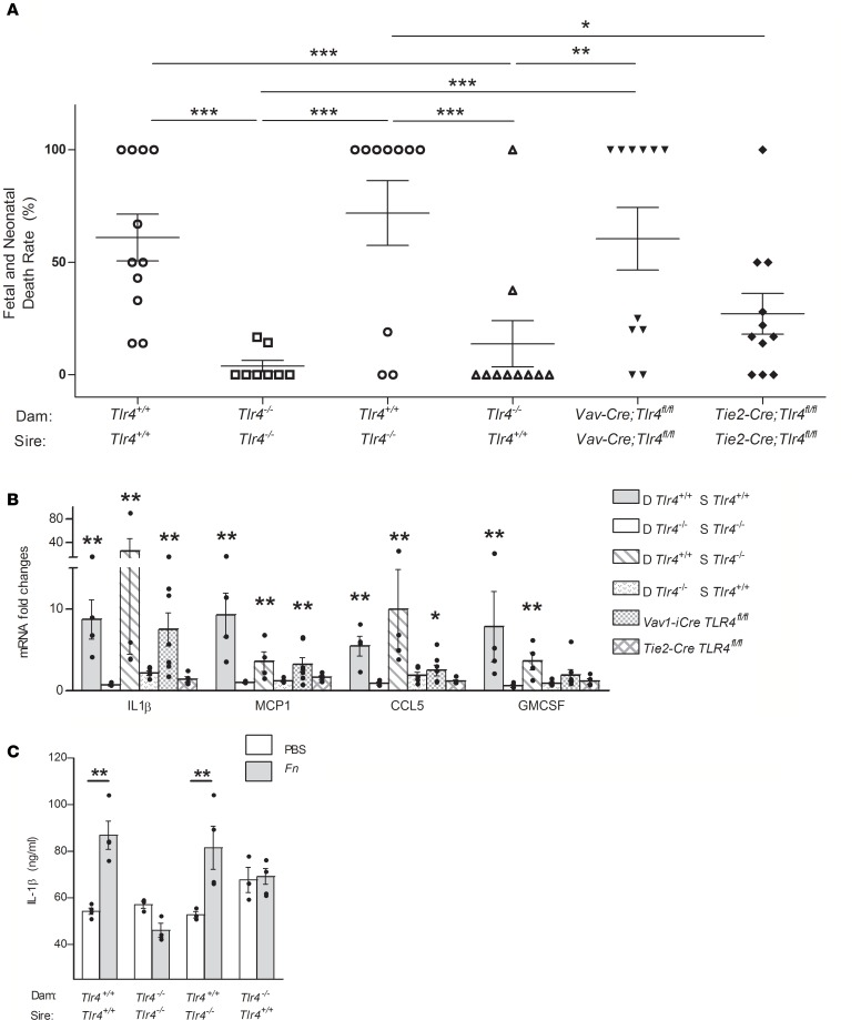
Figure 2. F.
nucleatum–induced placental inflammation originates from the maternal endothelial cells. Approximately 107 CFU F. nucleatum 12230 or saline was injected into the tail vein of each C57BL/6 pregnant dam on day 16 or 17 of gestation. (A) The fetal and neonatal death rate was calculated as the percentage of dead fetuses and neonates per litter at birth and followed up to 3 days after birth. Genotypes of the mating parents are shown below the x axis. Each geometric symbol represents 1 pregnant mouse. The horizontal lines indicate the average death rates. (B) mRNA levels in the placentas measured by real-time quantitative PCR and expressed as fold changes compared with each genotypes’ own saline-injected control groups. At least 3 pregnant dams were included in each group. (C) Protein levels of IL-1β in the placentas, as determined by ELISA. At least 3 pregnant dams were included in each group. For B and C, the placentas were collected at 48 hours after injection and pooled for each pregnant dam. The results are presented as dot plots with average and SEM. Two-way ANOVA was performed with simple-effects analysis when the interaction was significant. Student-Newman-Keuls was applied for post-hoc comparisons. *P < 0.05, **P < 0.01, ***P < 0.001. D, Dam; S, Sire.

