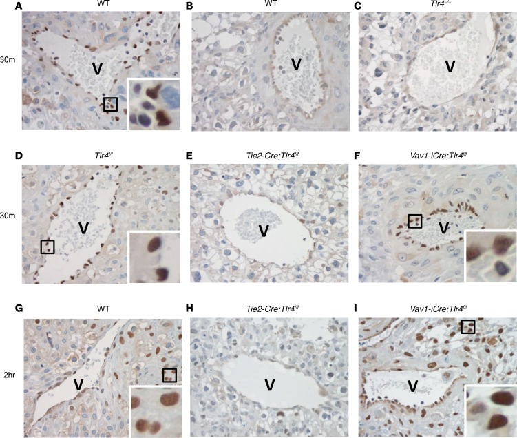
Figure 3. F. nucleatuminduces nuclear translocation of the p65 subunit of NF-κB in the decidua.
Immunohistochemical staining of the p65 subunit of NF-κB in the decidua of wild-type C57BL/6 (A, B, and G), Tlr4–/– (C), Tlr4f/f (D), Tie2-Cre;Tlr4f/f (E and H), and Vav1-iCre;Tlr4f/f (F and I) mice at 30 minutes (A–F) and 2 hours (G–I) after injection with saline (B) or approximately 107 CFU F. nucleatum (A, C, and D–I). Immunoreactive p65 was detected in the endothelial nuclei in A, D, and F and in the cells surrounding the venous sinuses (V) in G and I (insets). No nuclear p65 was detected in B, C, E, or H. Original magnification, ×500; zoom, 3.5- to 5-fold (insets). Representative image of at least 3 placentas from 1–2 dams of each group was selected.

