
FIGURE 1.
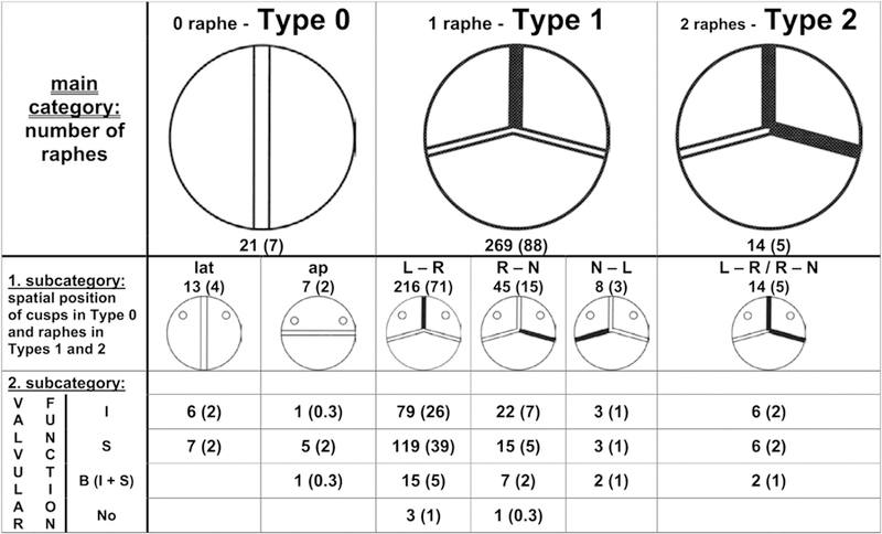
Sievers’ classification system for BAVas viewed from the surgeon’s side with the left coronary artery at left. The number of specimens is given, and the percentage is shown in parentheses. The blackened lines represent raphe. The main category is based on the number of raphes, the first subcategory is based on spatial position, and the second subcategory reflects valve function. Ap, Anterior-posterior; B, balanced valvular lesion; I, insufficiency; L, left coronary sinus; lat, lateral; N, noncoronary sinus; No, normal function; R, right coronary sinus; S, stenosis. Used with permission from Sievers and Schmidtke.14