
FIGURE 13.
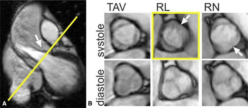
A, 2D balanced steady-state free precession cine MRI showing the left ventricular outflow tract and the position of the aortic valve imaging plane shown in yellow. B, TAV and the 2 most common BAV phenotypes: R/L and R/N cusp fusion. Arrows show the location of the raphe (if present) between the conjoined cusps. The conjoined R/L cusp (yellow box, arrow) is also seen to be doming in the corresponding left ventricular outflow tract view (A, arrow). Bicuspidality of the aortic valve should be assessed in systole rather than diastole, because the valves often appear tricuspid when closed. TAV, Tricuspid aortic valve; RL, right-left; RN, right noncoronary. Adapted from Entezari and colleagues.182