
FIGURE 14.
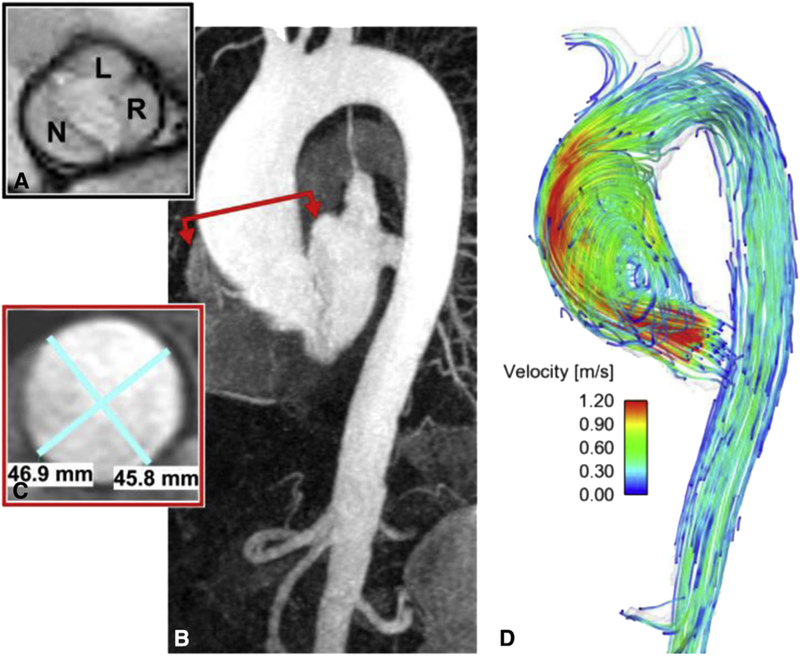
MRI of a 73-year-old man shows the (A) balanced steady-state free precession valve cines of a patient with BAV with R/L fusion and no stenosis. B, Contrast-enhanced magnetic resonance angiography shows mild dilation of the sinus of Valsalva with a maximal dimension of 40 mm and (C) a 47-mm dilation of the mid-ascending aorta. D, An eccentric jet is observed downstream from the nonstenotic bicuspid valve that impacts along the anterior portion of the tubular aorta. L, Left coronary cusp; N, noncoronary cusp; R, coronary cusp.