
FIGURE 6.
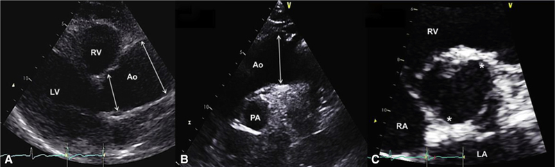
Typical echocardiographic findings in a patient with BAV with tubular ascending aorta dilatation phenotype. A, Echocardiogram of a 60-year-old woman with R/N BAV, no aortic valve regurgitation, and a fusiform ascending tubular aortic aneurysm. Left parasternal long-axis view in diastole shows root measurement of 36 mm (first arrow from left) and midtubular ascending aorta measurement of 47 mm (second arrow from left). B, Suprasternal diastolic view shows the mildly dilated proximal arch (36 mm, arrow) and normal upper descending aorta. C, Parasternal short-axis en face view of the aortic valve in systole shows 2 commissures (asterisks) at 1 and 7 o’clock with right nonfusion. LV, Left ventricle; RV, right ventricle; Ao, aorta; PA, pulmonary artery; RA, right atrium; LA, left atrium.