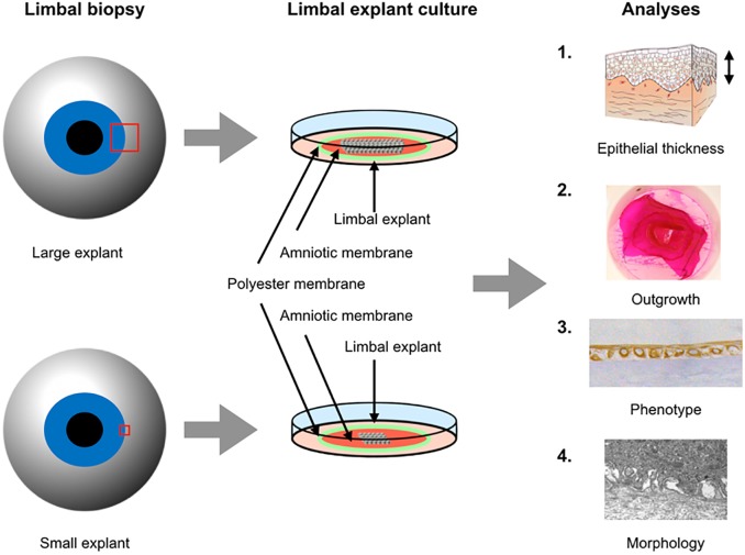
Fig 1. Experimental design of the study.
Large (3 mm) and small (1 mm) limbal explants were cultured for three weeks on intact amniotic membranes fastened to polyester membranes of culture plate inserts for 3 weeks. Epithelial thickness and stratification, outgrowth, phenotype and morphology were compared between the groups.

