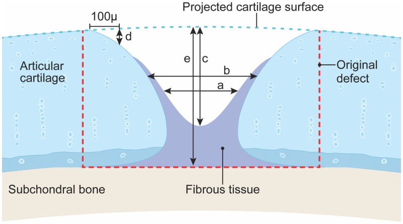
Fig 1. Histomorphometric measurements of lesion filling.
Five measurements were taken for lesion filling a) width of opening halfway between the lesion bottom and the projected original surface b) width, at the same depth as measurement a, of opening of physiological cartilage, c) depth of the opening at the center, d) depth at 100μm from edge of original lesion, and e) depth of original lesion, from the projected cartilage surface to the estimated original tidemark. Red dashed line represents the size of the original lesion (2.5 mm wide x ~ 1.5 mm deep).

