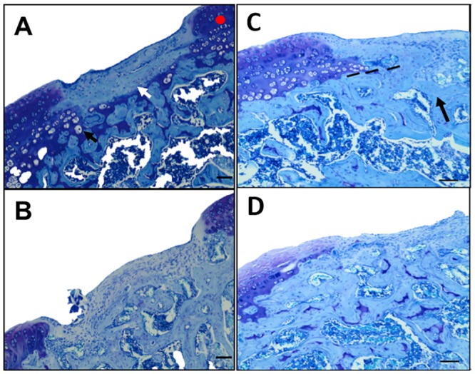
Fig 6. Lesion with sConstructs implants; Smooth fibrocartilage repair tissue, adjacent articular cartilage and subchondral bone repair.

Illustrative (median representative) photomicrographs (40x) of histochemical toluidine blue stained sections of rat knee lesions after 5 weeks of healing. Specimens selected for the photograph had the middle histology score for that group and thereby is a mid-representative of the differences. A) Lesions with BMP-2-sConstruct implants where the white arrow (⇨) points to smooth fibrocartilage repair tissue, the black arrow (➨) to chondrocyte/lacunae duplets, and the red dot, the histochemical staining for PG. B) Image as Fig 6A but from a control knee with suture only. C) Lesions with BMP-2-sConstruct implants where black arrow (➨) points to re-established subchondral bone and dotted line (- - - - -) is re-established tidemark, D) Similar image to Fig 6C from a control knee with suture only. Scale bar: 200 μm.
