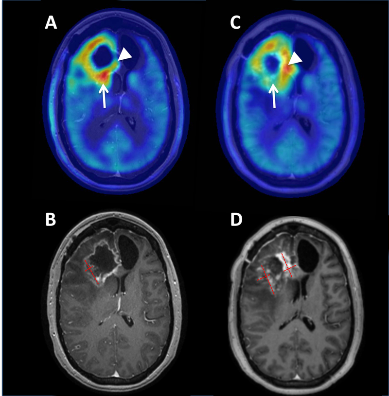
Figure 1.

Axial AMT-PET (A, C) and T1 post-contrast MR imaging (B, D) for patient 1. Pretreatment AMT-PET (A) and MRI (B) demonstrated an area of contrast enhancement with corresponding increased AMT uptake with maximum standardized uptake value (SUV) lesion/cortex ratio of 1.65 (arrow) in the posterior portion of the enhancing lesion, consistent with tumor recurrence. On-treatment AMT-PET (C) demonstrated an interval decrease of AMT uptake in this region with an SUV ratio of 1.48, suggesting that the observed expansion of MRI contrast enhancement (D) was pseudo-progression. However, other regions of the tumor such as the medial frontal component (arrowheads) demonstrated an interval increase of AMT uptake (from 1.38 to 2.10 tumor/cortex ratio) consistent with tumor progression in the expanding contrast enhancing mass. Red dashed lines demonstrate the measurements of the baseline lesion and the expanded contrast-enhancing masses on the follow-up MRI (with the sum of the areas increasing from 4.8 cm2 to 8.3 cm2).
