Figure 6:
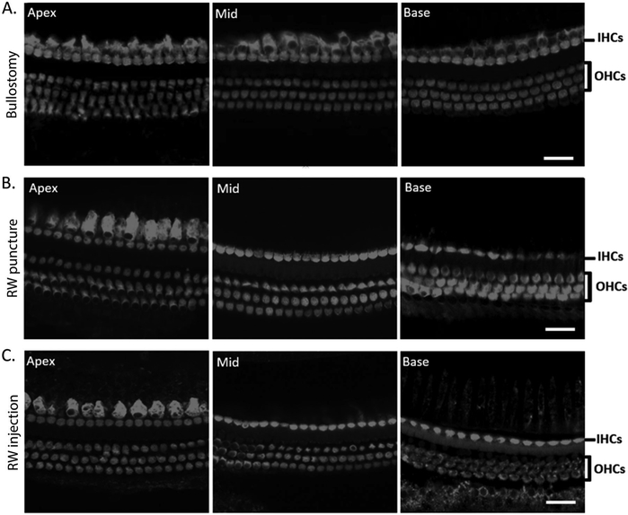
Hair cells are intact in mice with effusion and hearing loss after bullostomy, RW puncture, or RW injection. A. Representative images of a cochlea from a mouse with effusion following bullostomy alone. Inner hair cells (IHCs) and outer hair cells (OHCs) are intact in all three cochlear turns (scale bar = 20 μm). B. Representative images of a cochlea from a mouse with effusion following RW puncture. IHCs and OHCs are intact in all three cochlear turns (scale bar = 20 μm). C. Representative images of a cochlea from a mouse with effusion following RW injection. IHCs and OHCs are intact in all three cochlear turns (scale bar = 20 μm).

