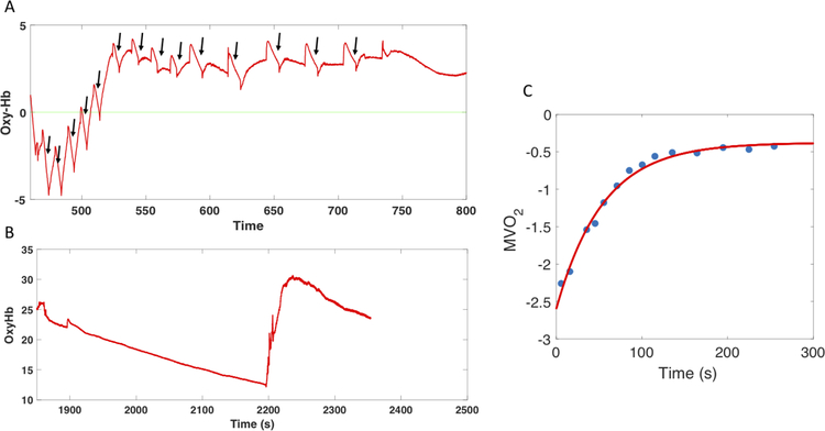
Figure 3. Use of Near Infrared Spectroscopy for the assessment of skeletal muscle oxygen consumption.
Panel A shows measurements of oxygenated hemoglobin during intermittent occlusions post-exercise (to calculate individual slopes, indicated by arrows). This signal is calibrated according to an ischemic occlusion (panel B). A subsequent exponential fit of the slopes (panel C) allows for the measurement of oxygen consumption (MVO2).

