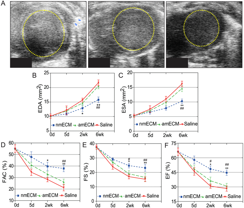
Figure 1. In vivo echocardiographic analysis of cardiac function.
(A) Short-axis B-mode echocardiography images of the left ventricle at diastole showing expansion of the left ventricle 6 weeks post-injury. Quantification of end diastolic (B) and systolic (C) area show reduced ventricle size in nmECM-treated mice. Cardiac function, shown as fractional area change (D), fractional shortening (E), and ejection fraction (F), is not preserved in saline or amECM-treated mice but significantly improved in nmECM-treated mice. (n = 8 per group; *p < 0.05, **p < 0.01 relative to saline; #p < 0.05, ## p < 0.01 relative to amECM).

