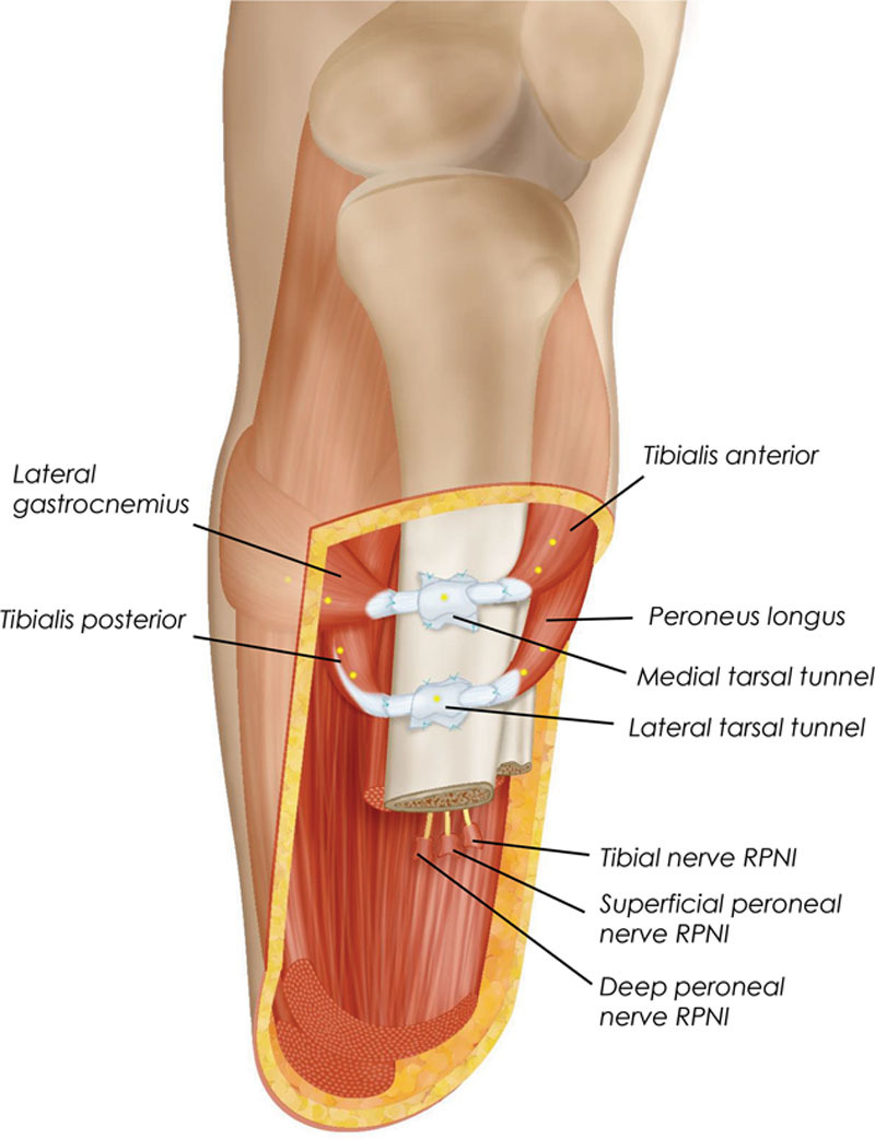
Fig. 4.

Schematic illustration of the Ewing amputation. Two AMIs are constructed in the residuum at the time of primary transtibial amputation. Tarsal tunnels harvested from the amputated ankle joint are affixed to the medial flat of the tibia and serve as pulleys for the AMIs. When the patient is connected to a robotic prosthesis, the proximal and distal AMIs are myoelectrically linked to the prosthetic ankle and subtalar joints, respectively. In the diagram, suture points are denoted by blue crosses and tantalum beads are denoted by yellow circles. Positioning of these elements are representative, and not to scale.
