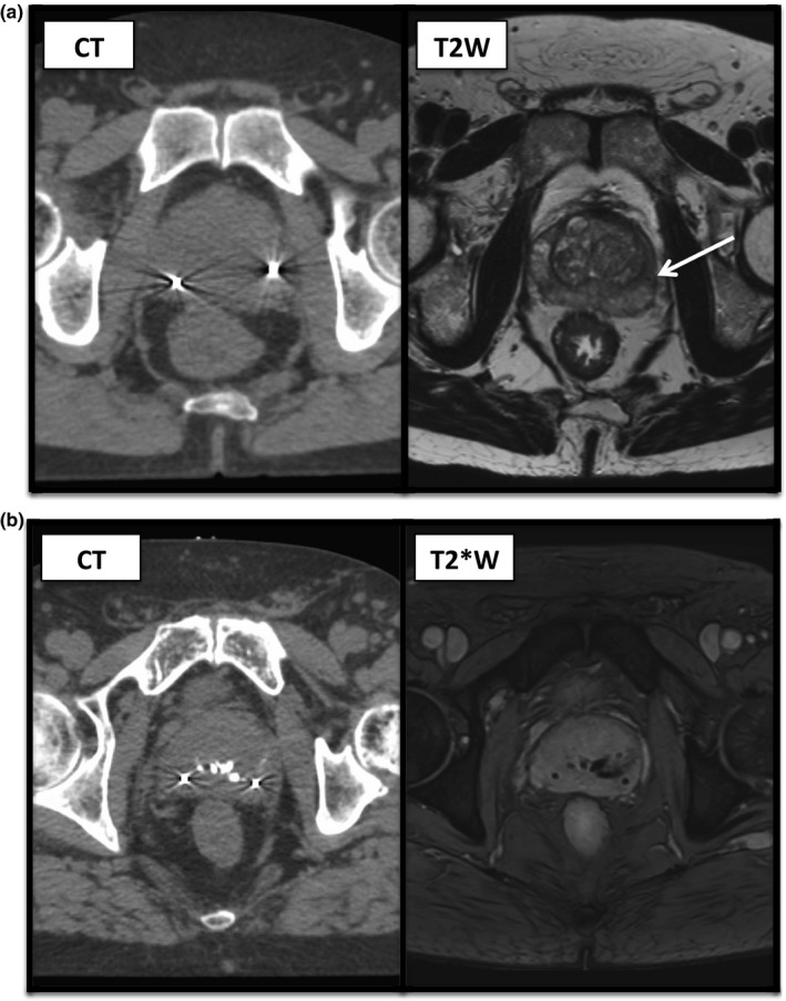
Figure 6.

(a) Corresponding CT (left) and T2W (right) images for a patient showing the appearance of a fiducial marker on standard T2W imaging, as indicated by the arrow. The second fiducial marker visible on CT imaging could not be identified on T2W images here. (b) Corresponding CT (left) and T2*W (right) images for a patients showing two fiducials with surrounding artifact on CT images and central calcifications, all showing as signal loss on T2*W imaging.
