. 2019 Mar 12;8:e41431. doi: 10.7554/eLife.41431

Figure 1. Antibody-free magnetic selection of HIV-infected primary T cells.

(A) Workflow for AFMACS-based magnetic selection of HIV-infected primary T cells. (B) Schematic of HIV-AFMACS provirus (pNL4-3-ΔEnv-Nef-P2A-SBP-ΔLNGFR). For simplicity, reading frames are drawn to match the HXB2 HIV-1 reference genome. Length is indicated in base pairs (bp). The complete sequence is available in Supplementary file 1. Nef-hu, codon-optimised Nef; RRE, Rev response element; SP, signal peptide. (C) Expression of cell surface SBP-∆LNGFR and CD4 on primary T cells 24 or 48 hr post-infection with HIV-AFMACS. Cells were stained with anti-LNGFR and anti-CD4 antibodies at the indicated time points and analysed by flow cytometry. (D–E) Magnetic sorting of HIV-infected (red, LNGFR+, CD4 low) and uninfected (blue, LNGFR-, CD4 high) cells. Cells were separated using AFMACS 48 hr post-infection with HIV-AFMACS and analysed as in (C). Representative (D) and summary (E) data from six independent experiments in CEM-T4s (triangles) and primary T cells (circles) are shown, with means and 95% confidence intervals (CIs).

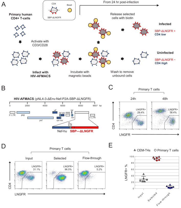
Figure 1.

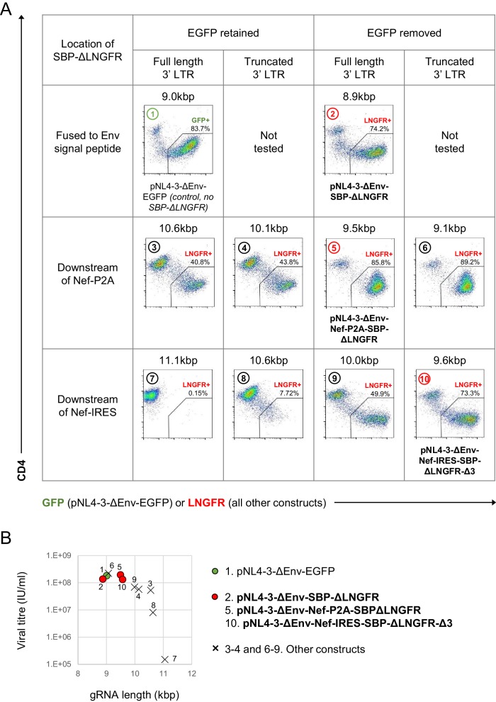
Figure 1—figure supplement 1. Initial screen of SBP-ΔLNGFR-expressing HIV viruses.

Figure 1—figure supplement 1.

(A–B) Expression of GFP (pNL4-3-ΔEnv-EGFP only) or cell surface SBP-∆LNGFR (all other constructs) and CD4 on CEM-T4s 48 hr post-infection with indicated pNL4-3-ΔEnv-based viruses (A). Cells were stained with anti-LNGFR and anti-CD4 antibodies and analysed by flow cytometry. gRNA length is shown for each construct, and compared with functional viral titre derived from the % LNGFR +cells (B). Each construct is numbered, and the three proviruses selected for further testing are highlighted (red, bold text). pNL4-3-ΔEnv-EGFP was included as a control (green).

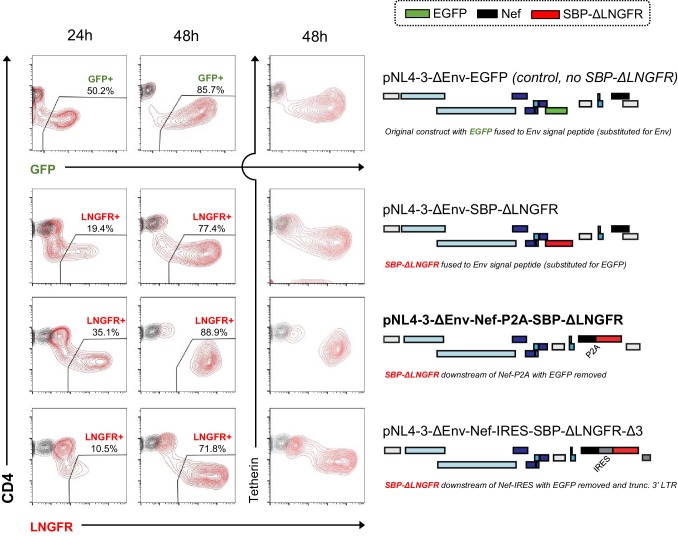
Figure 1—figure supplement 2. Time course evaluation of selected SBP-ΔLNGFR-expressing HIV viruses.

Figure 1—figure supplement 2.

Expression of GFP (pNL4-3-ΔEnv-EGFP only) or cell surface SBP-∆LNGFR (all other constructs) and CD4 or tetherin in CEM-T4s 24 or 48 hr post-infection with HIV-AFMACS. Cells were stained with anti-LNGFR and anti-CD4 or anti-tetherin antibodies at the indicated time points and analysed by flow cytometry (red, infected cells; grey, mock infected cells). Schematics indicate the location/setting of SBP-ΔLNGFR within each provirus, with ORFs and non-coding features coloured as in Figure 1B (but with Nef in black and EGFP in green). For simplicity, reading frames are drawn to match the HXB2 HIV-1 reference genome. The final HIV-AFMACS virus is highlighted (pNL4-3-ΔEnv-Nef-P2A-SBP-ΔLNGFR, bold text). pNL4-3-ΔEnv-EGFP was included as a control.