
Figure 9.
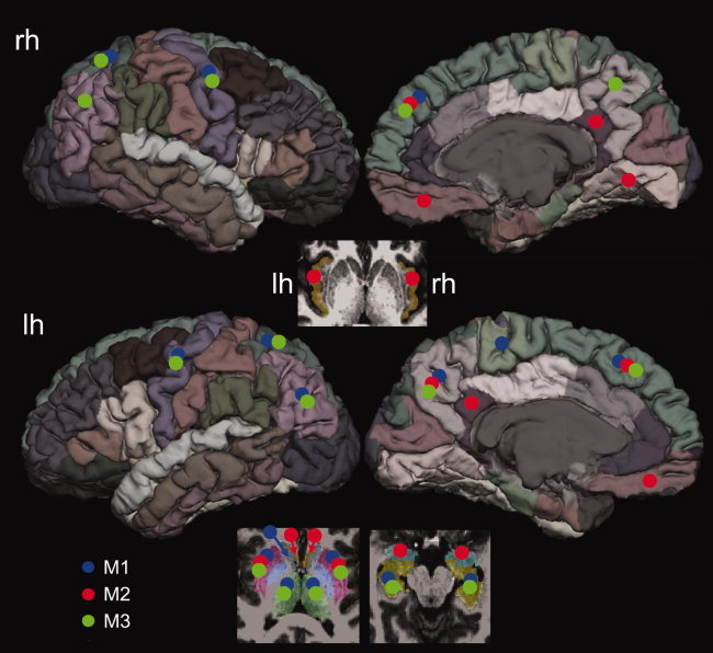
A summary of the top 20% nodes with the highest nodal BC values derived in the present study. The same diffusion MRI data, partitioning scheme were used in all three reconstruction methods. Large variability can be observed from the most central nodes (20%) based on the ranks of BC measures. The bilateral putamen, bilateral superior frontal lobes, and left precuneus were unanimously identified as hubs in all three methods. The positions of the marking circles are in the approximate locations. [Color figure can be viewed in the online issue, which is available at wileyonlinelibrary.com.]