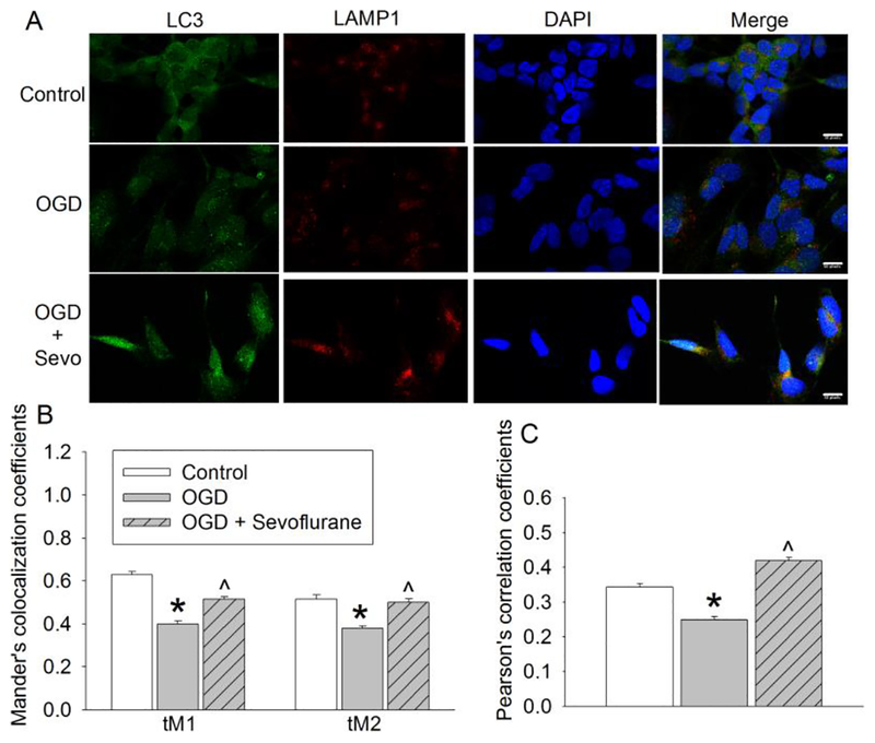
Fig. 4. Sevoflurane improved autophagosome-lysosome fusion.
Human neuron-like cells were subjected to 1 h OGD followed with 20 h simulated reperfusion. Cells were exposed to 2% sevoflurane for 1 h immediately after the onset of simulated reperfusion. A: Representative confocal microscopic images. Scale bar = 10 μm. B: Mander’s co-localization coefficients. C: Pearson’s correlation coefficients. Results are means ± S.E.M. (n = 150 cells). * P < 0.05 compared to control. ^ P < 0.05 compared to OGD only. OGD: oxygen-glucose deprivation

