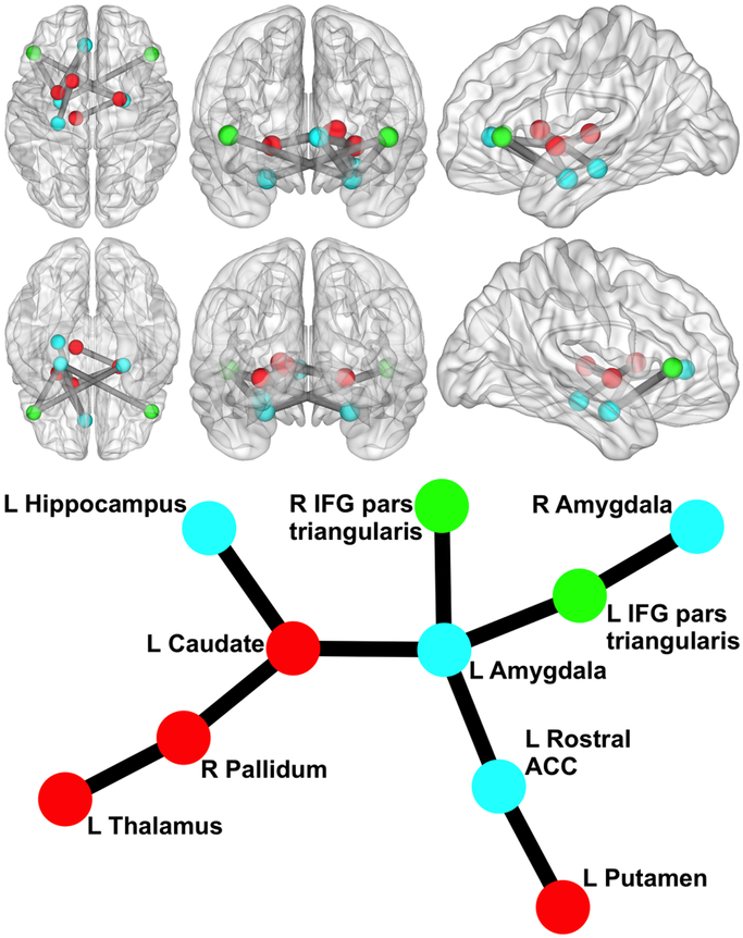
Figure 1. Network Related to Externalizing Polygenic Score.
Circle/sphere color reflects module. Stick/ball figure created using Kamada-Kawai spring embedder algorithm (SONIA; Bender-deMoll and McFarland, 2006). R = right; L = left; ACC = anterior cingulate cortex; IFG = inferior frontal gyrus. Node color indicates different modules. The six 3d brain images show (clockwise from top left) an axial view from superior to the brain, a coronal view from anterior to the brain, a sagittal view from left of the brain, a sagittal view from right of the brain, a coronal view from posterior to the brain, and an axial view from inferior to the brain (created via BrainNet Viewer; Xia et al., 2013).

